
Chiropractor Chandler AZ
PLATELET-RICH PLASMA PROLOTHERAPY FOR KNEE CARTILAGE DEGENERATION
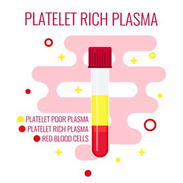
Our blood consists of 95% RBC, 1% WBC and 4% platelets. The role of platelets is blood clotting, but they have the ability to stimulate and accelerate the repair machinery of the body. This characteristic is manipulated in platelet-rich plasma Prolotherapy in which a person's own platelets are used to repair the damage in the body.
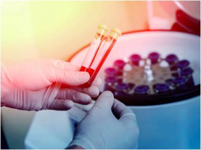
In PRP Prolotherapy, the blood of the patient is drawn under sterile conditions and centrifuged to separate platelet-rich plasma from other blood components. It is then injected back into the patient at the site of injury where it gets activated and releases alpha granules.
Alpha granules contain growth factors which are the proteins that initiate the tissue repair by cell growth and proliferation. Platelets cause the growth factors to stimulate the epithelial growth factors (EGF) which induce the cell migration and replication at the site of damage, causing the damaged tissues to heal quickly. The process of repair consists of three steps:
Inflammation phase: This lasts for 2-3 days. In this phase, growth factors are released.
Proliferation phase: This lasts for 2-4 weeks. It is vital for musculoskeletal regeneration.
Remodeling phase: This lasts over a year. In this phase, collagen is matured and strengthened.
PRP is very safe since the chances of blood-borne infection and immunogenic reactions are completely eliminated. The only risks involved are no pain relief, scar tissue formation, and neurovascular injury.
PLATELET-RICH PLASMA PROLOTHERAPY FOR KNEE CARTILAGE DEGENERATIONThe knee joint is the largest and most complex joint in the body. It not only bears the weight of the body, but also helps to perform basic functions of the daily life such as walking, running and standing. Due to increased use of the knee joint, it is most susceptible to traumas and pain. One of the major causes of knee pain is cartilage injury and degeneration. 20-70% of knee pain is caused by articular cartilage degeneration and trauma. Each year, over two million Americans suffer from cartilage related pain.
Injury to knee cartilage can disturb the normal load of the joint. This injury can occur due to trauma or just normal daily activities. The disturbance of the delicate knee balance leads to overload knee damage. With the passage of time, the initial small defect in the articular cartilage or tear in the meniscal cartilage can progress. This gradual damage can lead to degenerative joint disease.

The knee joint is made up of three bones, two different types of cartilages, and four major ligament groups.
Bones : The knee joint is made up of three bones: Femur, Tibia, and Patella.
Cartilages : The end of Femur, Tibia and Patella are covered by articular (Hyaline) cartilage. It is a white substance that has the consistency of strong rubber, but is actually a mixture of collagen and special large sponge-like molecules. This structure is maintained by living cells called chondrocytes. To allow the normal and smooth joint motion, the surface of the articular joint is covered with fluid which makes it very slippery.
The second type of knee cartilage is called meniscal Fibrocartilage. This cartilage is present in the form of C-shaped pads that are located between the Tibia and Femur. One meniscus cartilage is present on each side of the knee. The medial meniscus is present on the inner thigh and the lateral meniscus is present on the outer side of the thigh. They protect the articular cartilage by acting as a shock absorber and distributing joint force.
Ligaments : These are rope-like structures that connect two bones. Four main ligaments are present in the knee: two inner cruciate ligaments and two outer collateral ligaments. These ligaments stabilize the knee joint and protect the articular cartilage and menisci from trauma.
CAUSES OF KNEE CARTILAGE DEGENERATIONThere are several causes of knee cartilage degeneration. Some of the important causes are the following:
Knee Trauma: The most obvious initiating factor of knee cartilage degeneration is a direct trauma to the knee. This can be in the form of a single incident or multiple micro-traumatic events over a prolonged period.

Obesity: Obesity is a risk factor for knee cartilage degeneration. Increased weight is associated with osteoarthritis as well. Studies have shown that weight loss is directly related to the reduced chances of cartilage degeneration. In the Framingham study, the risk of knee degeneration was reduced by 50% in women who lost an average of 5 kg (11 lbs).
Meniscal tear: A meniscal tear is the most common cause of knee cartilage degeneration leading to surgery in America. It is caused by daily activities, with or without trauma. The symptoms of a meniscal tear are pain and swelling. The meniscal tear causes the body weight to be distributed unevenly which then leads to articular cartilage damage.
Ligament damage: Ligaments are present in the knee joint to stabilize its structure. Any damage to ligaments leads to knee joint instability. As a result, the knee joint moves around too much which slowly damages the knee cartilage. Signs and symptoms of ligament damage are soreness or swelling after physical activity.
Medication: Medications such as local anesthesia and steroid injections cause cartilage cell death. In addition, commonly used NSAID medications like Ibuprofen, Naproxen, and Celebrex may also have adverse impacts on normal cartilage cells.
Osteoarthritis: Osteoarthritis is a condition in which the knee cartilage wears away. It is the most common type of arthritis to affect women more than men and most likely develops after the age of 45. According to the Arthritis foundation, more than 45 million Americans have osteoarthritis. Risk factors are age, gender, heredity, trauma, and other diseases can also trigger osteoarthritis.
SIGNS AND SYMPTOMS OF KNEE CARTILAGE DEGENERATIONPatients with cartilage degeneration experience the following signs and symptoms:
Inflammation: The knee joint swells, becomes warmer than the rest of the body and becomes painful.
Stiffness: The knee joint becomes stiff.
Limitation in movement: As the damage progresses, the motion range of the knee is reduced.

Early detection of knee cartilage degeneration is important before it progresses and fully depletes the cartilage. Diagnosis of cartilage degeneration is difficult and unreliable and Radiography and MRI provide low sensitivity and inadequate diagnostic precision. Some of the ways that are used to diagnose cartilage degeneration include:
Arthroscopic examination: It is an invasive method that requires anesthesia, but it is helpful to diagnose the cartilage problem. It probes the articular cartilage surface while making a video which can be used later to diagnose cartilage degeneration.
Magnetic resonance Imaging (MRI): A standard clinical MRI cannot diagnose the articular cartilage defects, but the advanced MRI techniques and special articular cartilage scanning protocols have increased the accuracy of diagnosis.
TREATMENT OF KNEE CARTILAGE DEGENERATIONConservative treatment methods include non-steroidal anti-inflammatory drugs, physical therapy, and pain killers. Some patients respond well to the conservative treatment methods, for others, surgery is performed.
PLATELET-RICH PLASMA THERAPY FOR KNEE CARTILAGE DEGENERATIONConventional treatment methods such as physical therapy, exercises, painkillers and NSAIDs are helpful in symptom management and improving mobility, but they do not repair the cartilage.
Chondrocytes, the cells that synthesize articular cartilage, are active cells that have the ability to divide and produce cartilage. In normal cartilage, this process is strictly regulated, but in knee degeneration, this process is imbalanced and the messenger molecules, called cytokines, increase the cartilage breakdown more than cartilage repair.
To treat the cartilage degeneration, the most suitable treatment option is the one that increases the chondrocytes activity, cartilage repair, and regulates cytokine production. This can be achieved by platelet-rich plasma (PRP) Prolotherapy. A chronic knee pain study published in the Journal of Prolotherapy showed amazing pain-relieving effects of PRP Prolotherapy in patients with knee cartilage degeneration.
PRP Prolotherapy improves overall joint homeostasis, reduces synovial membrane hyperplasia, and moderates the cytokine level, thus leading to improved cartilage condition, without affecting the cartilage structure.
In 2013, a study was done on 78 patients with osteoarthritis in both knees. Each knee received either 1 PRP injection, 2 PRP injections, or 1 placebo saline injection. The knees were evaluated after 6 weeks, 3 months, and 6 months. Results showed pain reduction in knees that received 1 or 2 PRP injections at 6 weeks and 3 months. At the 6th month check, the positive results declined although the pain and knee function was still better. The group that received placebo injections showed no positive results and pain and stiffness was increased in their knees.
Not all clinical studies show that PRP Prolotherapy improves the knee cartilage degeneration. In some cases, the results were not different from the placebo treatments. It is due to the difference in procedure and PRP formulation. Therefore, it is important to design a procedure that is effective and efficient which helps the patients with knee cartilage degeneration so that they can live a healthy, active life.
Conservative Treatments to Combine with PRPWhile PRP and stem cell treatments are enhancing the tissue repair and regeneration, conservative treatments can enhance healing, strengthen the muscles, and stabilize joint movements to maximize your recovery.
Cold Laser Therapy Treatments
- ACCELERATED TISSUE REPAIR AND CELL GROWTH
- FASTER WOUND HEALING
- REDUCED FIBROUS TISSUE FORMATION
- ANTI-INFLAMMATION
- PAIN RELIEF
- INCREASED BLOOD FLOW
- INCREASED REPAIR AND REGENERATION
- NERVE FUNCTION AND REPAIR
- INCREASED ENERGY PRODUCTION - ATP
Photons of light from lasers penetrate into tissue and accelerate cellular growth and reproduction. Laser therapy increases the energy available to the cell so it can work faster, better, and quickly get rid of waste products. When cells of tendons, ligaments, and muscles are exposed to laser light they repair and heal faster.

Laser light increases collagen production by stimulating fibroblasts. Collagen is the building block of tissue repair and healing. Laser therapy increases fibroblast activity and therefore collagen production to speed healing.
Low level laser therapy decreases scar tissue formation. Scar tissue can be a source of chronic pain and poor healing. By eliminating excessive scar tissue and encouraging proper collagen production, painful scars and chronic pain is reduced.
Laser therapy causes vasodilatation (increases size of capillaries) which increases blood flow. The treatments also increases lymphatic drainage to decrease swelling or edema. Therefore, laser therapy reduces swelling caused by bruising or inflammation while speeding the recovery process.
Cold laser therapy decreases pain by blocking pain signals to the brain. Some nerve cells sense pain and send signals to the brain. Chronic pain can be caused by overly active pain nerves. Specific wavelengths help "shut off" the pain signals, thereby eliminating your pain.
Low level lasers are excellent at decreasing inflammation, which also increases pain nerve activity. Cold laser therapy also increases endorphins and enkephalins, which block pain signals and decreases pain sensations. Overall, laser therapy reduces painful nerve signals and reduces your perceived pain.
Blood carries nutrients and building blocks to the tissue while carrying waste products away. Increased blood flow to tissues increases and enhances cellular healing. Cold laser therapy increases the formation of capillaries in damaged tissues. Specific laser frequency also increases blood flow to the area treated to enhance injury repair.
Low level lasers increase enzyme activity to improve metabolic activity which affects cell repair and regeneration. The enzymes are turned on "high" to speed the healing.
Nerves heal very slowly. Lasers speed up this process. Damage to nerves causes numbness, pain, muscle weakness, and altered sensations. Laser therapy treatments enhance nerve function, healing, and reduce pain.
ATP is like gasoline for cells, it is the energy source that cells operate. Injured cells often have low levels of ATP, which decreases their ability to heal and repair. By increasing ATP and "gasoline storage levels," cells have more ability to heal and repair.
Therapeutic treatments for addressing soft tissue injuries involve massage therapy, manual therapy, trigger point therapy, Graston Technique, or Active Release Technique. These treatments increase blood flow, decrease muscle spasms, enhance flexibility, speed healing, and promote proper tissue repair.
When these treatments are incorporated into a treatment plan, patients heal faster and are less likely to have long-term pain, soft tissue fibrosis, or scar tissue in the injured muscle. These soft tissue treatments are incorporated with therapeutic exercises and flexibility programs.
Many leg injuries are associated with radiating pain. The two legs function as a system for movement. Injuries in one area of the system are commonly associated with poor joint stabilization in the foot, knee, or hip. This leads to poor alignment and excessive forces being placed onto muscles and tendons. Knee injuries are common because of weakness and poor stabilization of the leg and hip muscles. The combination of muscle weakness, poor coordination, and altered gait mechanics produce excessive strain on the soft tissues.

The lower extremity works as a comprehensive unit performing many of the repetitive tasks at home, work, and recreational sports. Injuries to one area of the musculature often indicate that additional damage has been incurred by other muscles.
Many therapeutic exercises can help restore proper strength and endurance to the leg muscles. Isometric exercises are often the initial treatment exercises. Followed by single plane rubber band exercises for hip, knee, and ankle; flexion, extension, adduction, abduction, circumduction, inversion, and eversion. Dynamic exercises involving stability foam, rubber discs, exercise balls, and BOSU balls can be performed on the floor. The more unstable the surface, the more effort and stabilization is required of all the lower extremity muscles.
Vibration plates enhance neuromuscular learning throughout the ankle, knee, foot, hip, and back muscles. Additional strength exercises can be found on the hip, knee, and foot strengthening pages. More information for injuries and treatments foot pain and exercises.
Bibliography:
Heidari, B. (2011). Knee osteoarthritis prevalence, risk factors, pathogenesis and features: Part I. Caspian J Intern Med, 205–212.
Litwic, A., Edwards, M., Dennison, E., & Cooper, C. (2013). Epidemiology and Burden of Osteoarthritis. Br Med Bull, 185–199.
Raeissadat, S. A., Rayegani, S. M., Babaee, M., & Ghorbani, E. (2013). The Effect of Platelet-Rich Plasma on Pain, Function, and Quality of Life of Patients with Knee Osteoarthritis. Pain Research and Treatment, doi:10.1155/2013/165967.