
Chiropractor Chandler AZ
PLATELET-RICH PLASMA PROLOTHERAPY FOR KNEE ARTHRITIS
Knee arthritis is a degenerative condition that affects millions of people around the world. It is an inflammation of the knee joint. Although arthritis can affect any joint in the body, the knee joint is the most susceptible joint to arthritis.
Knee arthritis affects the whole life of the patient, making it difficult to perform basic daily functions on his own. It makes activities like walking, standing, and running difficult. More than 100 different types of arthritis have been diagnosed and the most common types are osteoarthritis and rheumatoid arthritis.
In 2012, more than 52 million people were diagnosed with arthritis according to the National Health Interview Survey. Women are more affected with knee arthritis than men. The larger portion of arthritic patients are ageing people, especially those who are above the age of 60.

The knee is the largest and strongest joint in the human body. It is a synovial joint, made up of bones, cartilages, tendons and ligaments. Its function is to provide mobility to the lower leg.
BONES: The knee joint is made up of three bones: Femur, Tibia, and Patella.
ARTICULAR CARTILAGE: The ends of the femur and Tibia and the back of Patella are covered by articular cartilage.
MENISCUS: Two wedge-shaped pieces of meniscal cartilage act as "shock absorbers" between the femur and tibia.
LIGAMENTS: There are four knee ligaments that bind the bones in the knee joint to each other. These ligaments are divided into two groups: two Collateral Ligaments and two Cruciate Ligaments.
TENDONS: Tendons connect muscle to the bone. Quadriceps tendon, also called patellar ligament, is a major support for the knee joint. The Patella bone lies within this tendon.
SYNOVIAL SHEATH: The inner surface of the joint is lined by a thin synovial membrane except where there is articulate cartilage. This produces synovial fluid which lubricates the joint cavity.
CLASSIFICATION OF ARTHRITISThere are more than 100 types of arthritis. The most common types are:
Rheumatoid Arthritis:It is an autoimmune disease that affects multiple joints within the body. In this condition, both of the knee joints are affected simultaneously. The synovial membrane that acts as a cushion in the knee joints is swollen which causes pain and discomfort. The ligaments, tendons, and bones in the knee joint are attacked by the immune cells that weaken them as the disease prolongs. If this condition is not managed properly, it leads to rheumatoid arthritis.
Osteoarthritis:The most common type of arthritis that affects people of age 50 and above is osteoarthritis. It is a degenerative condition which, unlike rheumatoid arthritis, develops when the cartilage of the knee gradually deteriorates. Due to the absence of cartilage, the friction between the bones increases which causes the pain and leads to more damage with the passage of time. The development of osteoarthritis is slow and the pain gets worse with time.
Posttraumatic arthritis:Any injury to the knee can lead to the development of arthritis later in life. A broken bone, ligament, or tendon puts an extra burden on the knee that can lead to the development of arthritis later in life.
SYMPTOMS OF KNEE ARTHRITIS:- Pain in the knee
- Inflamed knee
- Increased stiffness in the knee that makes bending of knee difficult
- Increased knee pain in the morning
- Increased knee pain after sitting for a long span of time
- Increased knee pain after intense physical activity
- Knee lock

DIAGNOSIS OF KNEE ARTHRITIS
Physical examinationDuring the physical examination, the doctor will look for the following signs to diagnose the knee arthritis:
- Knee joint swelling and redness around the knee
- Knee joint tenderness
- Pain when pressure is exerted on the knee
- Other arthritis symptoms
A detailed picture of the knee joint structure is created by using an X-ray. The narrowing of space between the joint, changes in the bone, and osteophytes formation are the signs of an arthritic knee.
MRI, computed tomography, or a bone scan are helpful in determining the bone condition of the knee.
Blood testA blood test is helpful in the diagnosis of rheumatoid arthritis.
TREATMENT FOR KNEE ARTHRITISArthritis cannot be treated, only its symptoms can be managed to relieve pain and avoid disability.
Nonsurgical treatmentFirst, the knee arthritis is treated by nonsurgical treatment options. The following are some of the conventional treatment options for knee arthritis:
Lifestyle modificationIt includes basic changes such as reducing high-intensity activities, losing weight, and resting. These changes help to reduce pain in the knee.
Physical therapyPhysical therapy helps in increasing the range of motion and strengthens the muscles in the knee joint.

Other options include:
- Assistive devices such as a cane
- Wearing shock absorber shoes
- Using ice, heat, and pain-relieving ointments
Painkillers and anti-inflammatory medicines are prescribed to patients with knee arthritis. NSAIDs, such as ibuprofen and naproxen, are also prescribed for knee arthritis pain. In the case of extreme pain, corticosteroid injection is injected into the arthritic knee to relieve the pain.
Surgical treatmentIf the above mentioned methods fail to relieve the pain and the situation gets worse, surgery is the only option left to make the knee condition better. There are different surgical treatments for a different type of complication:
Cartilage graftingIf the cartilage of the knee is completely worn out, the healthy cartilage from the other knee is used to fill the hole in the arthritic knee. Cartilage grafting is suitable for younger patients only who have less damaged knee joints. It is not suitable for rheumatoid arthritis patients where both the knees are affected.
SynovectomyIn the rheumatoid arthritis knee, the synovectomy is performed in which the inflamed synovial membrane is removed to relieve the pain.
ArthroplastyIn arthroplasty, the worn out piece of cartilage is replaced with a metal or plastic structure to restore the normal structure of the knee.
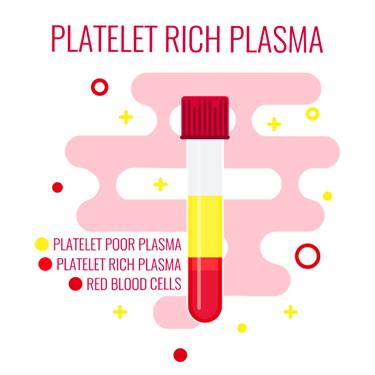
Arthritis is characterised by wear and tear of cartilage, bone, ligament, and tendons in the knee joint. The conventional treatment options either relieve the pain by manipulating the pain-signalling pathways of the body, removing the damaged part, or replacing it. None of the conventional methods regenerates the damaged structure. Therefore, a method that can regenerate the worn out structure in the arthritic knee is very helpful in knee arthritis. An alternative treatment option known as Platelet-Rich Plasma Prolotherapy is known to promote the regenerative properties of the body to repair the damaged part. It is a type of Prolotherapy that uses the platelet-rich plasma derived from the patient's blood to initiate and accelerate the self-healing at the site of injury.
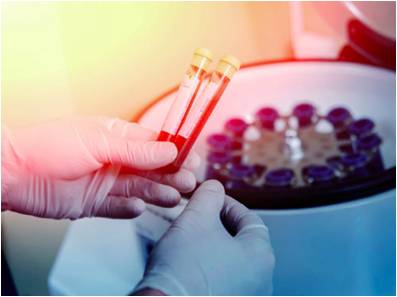
In platelet-rich plasma (PRP) Prolotherapy, the blood is drawn from the patient under sterile conditions. The blood is then centrifuged to separate the platelet-rich plasma from rest of the components. It is then injected back at the site of injury. The major role of platelets is in blood clotting, but they contain alpha particles that are rich in growth factors, thus platelets also play an important role in tissue repair. After the PRP is injected into the body, it goes through following stages to repair the damage:
- INFLAMMATION PHASE that lasts for 2-3 days. In this phase, growth factors are released
- PROLIFERATION PHASE that lasts for 2-4 weeks. It is vital for musculoskeletal regeneration.
- REMODELLING which lasts over a year. In this phase, collagen is matured and strengthened.
Side effects associated with this method are minimal. There is no risk of blood-borne disease transfer and allergic reaction. The only risks involved are an infection, no relief of pain, neurovascular injury, and scar tissue formation. The loss of limb or death is rare, but possible.
PLATELET-RICH PLASMA PROLOTHERAPY FOR KNEE ARTHRITISUse of PRP prolotherapy for knee arthritis is widely evaluated by researchers. The data available shows that PRP Prolotherapy is not only helpful in relieving the pain but also in preventing arthritis from getting worse.
Mode of mechanism in PRP Prolotherapy is repairing the damaged part, relieving the pain, and restoring the normal structure of the joint. Theoretically, it is an ideal treatment option for knee arthritis as it not only restores the natural structure of the knee, but also prevents arthritis from more damage.
A research study has shown significant functional betterment in patients with knee cartilage pathology and its effect last at least 12 months.
A study performed in 2013 showed that PRP injection might slow the progression or worsening of osteoarthritis. The average arthritic knee can lose up to 5% of cartilage per year. The same study showed that there was no further cartilage damage in knee arthritis in more than 70% of the patients after one year of PRP injections.
When the PPR was injected in the arthritic knee, the patients who were resistant to conventional treatments showed improvements in pain and stiffness, thus improving the quality of life.
The available research data has also shown that PRP injection is more effective in early stages of arthritis. Although many research studies are available that support PRP Prolotherapy, this data is not enough. More research is required to establish the effectiveness of PRP Prolotherapy to get it approved from the FDA and supported by insurance companies so that more people can enjoy the benefit of this revolutionary method.
CONSERVATIVE TREATMENTS TO COMBINE WITH PRPWhile PRP and stem cell treatments are enhancing the tissue repair and regeneration, conservative treatments can enhance healing, strengthen the muscles, and stabilize joint movements to maximize your recovery.

COLD LASER THERAPY TREATMENTS
- Accelerated tissue repair and cell growth
- Faster wound healing
- Reduced fibrous tissue formation
- Anti-inflammation
- Pain relief
- Increased blood flow
- Increased repair and regeneration
- Nerve function and repair
- Increased energy production- ATP
Photons of light from lasers penetrate into tissue and accelerate cellular growth and reproduction. Laser therapy increases the energy available to the cell so it can work faster, better, and quickly get rid of waste products. When cells of tendons, ligaments, and muscles are exposed to laser light they repair and heal faster.
Laser light increases collagen production by stimulating fibroblasts. Collagen is the building block of tissue repair and healing. Laser therapy increases fibroblast activity and therefore collagen production to speed healing.
Low-level laser therapy decreases scar tissue formation. Scar tissue can be a source of chronic pain and poor healing. By eliminating excessive scar tissue and encouraging proper collagen production, painful scars and chronic pain is reduced.
Laser therapy causes vasodilatation (increases the size of capillaries) which increases blood flow. The treatments also increases lymphatic drainage to decrease swelling or edema. Therefore, laser therapy reduces swelling caused by bruising or inflammation while speeding the recovery process.
Cold laser therapy decreases pain by blocking pain signals to the brain. Some nerve cells sense pain and send signals to the brain. Chronic pain can be caused by overly active pain nerves. Specific wavelengths help "shut off" the pain signals, thereby eliminating your pain.
Low-level lasers are excellent at decreasing inflammation, which also increases pain nerve activity. Cold laser therapy also increases endorphins and enkephalins, which block pain signals and decrease pain sensation. Overall laser therapy reduces painful nerve signals and reduces your perceived pain.
Blood carries nutrients and building blocks to the tissue, and carries waste products away. Increased blood flow to tissues increases and enhances cellular healing. Cold laser therapy increases the formation of capillaries in damaged tissue. Specific laser frequency also increases blood flow to the area treated to enhance injury repair.
Low-level lasers increases enzyme activity to improve metabolic activity that affects cell repair and regeneration. The enzymes are turned on "high" to speed the healing.
Nerves heal very slowly. Lasers speed up this process. Damage to nerves causes numbness, pain, muscle weakness, and altered sensations. Laser therapy treatments enhance nerve function, healing, and reduce pain.
ATP is like gasoline for cells, it is the energy source that cells operate. Injured cells often have low levels of ATP, which decreases their ability to heal and repair. By increasing ATP and "gasoline storage levels," cells have the ability to heal and repair.
Therapeutic treatments for addressing soft tissue injuries involve massage therapy, manual therapy, trigger point therapy, Graston Technique, or Active Release Technique. These treatments increase blood flow, decrease muscle spasms, enhance flexibility, speed healing, and promote proper tissue repair.
When these treatments are incorporated into a treatment plan, patients heal faster and are less likely to have long-term pain, soft tissue fibrosis, or scar tissue in the injured muscle. These soft tissue treatments are incorporated with therapeutic exercises and flexibility programs.
Many leg injuries are associated with radiating pain. The two legs function as a system for movement. Injuries in one area of the system are commonly associated with poor joint stabilization in the foot, knee, or hip. This leads to poor alignment and excessive forces being placed onto muscles and tendons. Knee injuries are common because of weakness and poor stabilization of the leg and hip muscles. The combination of muscle weakness, poor coordination, and altered gait mechanics produce excessive strain on the soft tissues.
The lower extremities work as a comprehensive unit performing many of the repetitive tasks at home, work, and recreational sports. Injuries to one area of the musculature often indicate that additional damage has been incurred by other muscles.

Many therapeutic exercises can help restore proper strength and endurance to the leg muscles. Isometric exercises are often the initial treatment exercises, followed by single plane rubber band exercises for hip, knee, and ankle; flexion, extension, adduction, abduction, circumduction, inversion, and eversion. Dynamic exercises involving stability foam, rubber discs, exercise balls, and BOSU balls can be performed on the floor. The more unstable of the surface the more effort and stabilization is required of all the lower extremity muscles.
Vibration plates enhance neuromuscular learning throughout the ankle, knee, foot, hip, and back muscles. Additional strength exercises can be found on the hip, knee, and foot strengthening pages. More information for injuries and treatments foot pain and exercises.
BIBLIOGRAPHY:
Heidari, B. (2011 ). Knee osteoarthritis prevalence, risk factors, pathogenesis and features: Part I. Caspian J Intern Med, 205–212.
Jang, S. J., Kim, J. D., & Cha, S. S. (2013). Platelet-rich plasma (PRP) injections as an effective treatment for early osteoarthritis. European Journal of Orthopaedic Surgery & Traumatology, 573–580.
Kamat, Y. D., Patel, N. G., Galea, A., Ware, H. E., & Dowd, G. S. (2014). Platelet-rich plasma injections for knee pathologies: a review. European Orthopaedics and Traumatology, 341–347.