Chiropractor Chandler AZ
PLATELET-RICH PLASMA PROLOTHERAPY FOR FOOT PAIN
Around 25% of human body's bones are found in the feet. There are 26 bones in each foot, 33 joints and more than 100 muscles, tendons and ligaments. Feet carry the weight of the whole body which makes them more prone to injuries.
In the United States, over 25,000 foot injuries are reported every year. The prevalence of foot injuries has also increased due to increasing popularity of competitive sports. In children, most of the foot injuries are sports related. The risk of a foot injury is higher in sports that involve jumping or a sudden change in direction. Dancers, gymnasts, soccer or basketball players are at a higher risk of having a foot injury.
In the older population, the risk of a foot injury is also high because they lose muscle mass and bone strength. Older people have vision and balance problems as well which increases the chance of a foot injury.

The foot is a complex structure of bones, joints, muscles and soft tissues. It is divided into three sections:
- Forefoot: It contains five toes (Phalanges) and five long bones (metatarsals)
- Midfoot: It is a pyramid-like assortment of bones that makes the curves of the foot. It includes three cuneiform bones, cuboid bone, and navicular bone.
- Hindfoot: It forms the heel and the ankle. The heel bone is the largest bone in the foot.
The complex movements of the feet are made possible due to muscles, tendons, and ligaments that run along the foot surface. The heel is connected to the calf muscles through the Achilles' tendons. These tendons are important for running, jumping, and standing.
CAUSES OF FOOT PAINPlantar Fasciitis: It is the inflammation of the plantar fascia ligament. Its symptoms are pain in the heel and arch that gets worse in the morning.
Osteoarthritis: The cartilage in the feet is worn out due to age and wear and tear. Symptoms of arthritis are pain, swelling, and feet deformity.
Gout: It is an inflammatory condition in which crystals are deposited in the joint. Severe pain and swelling are the symptoms. It affects the big toe most often.
Rheumatoid arthritis: It is an autoimmune condition that causes inflammation and joint damage. Rheumatoid arthritis affects joints in the feet, ankle, and toes.
Achilles tendon injury: The symptom of an Achilles tendon injury is pain in the back of the heel. Injury can be sudden or due to tendinitis.
Calluses: It is a build-up of tough skin on the foot. It usually forms on the balls of the foot or the heels and may be painful.
Heel spurs: It is an abnormal growth of bone in the heel. It may cause severe pain during walking or standing. The risk of developing heel spurs is higher in people with plantar fasciitis, flat feet, or high arches.
Ingrown toenails: In this condition, one or both sides of the toenail may grow into the skin. Ingrown toenails are painful and can lead to foot infections.
Sand Toe - Sprain of the big toe, where the toe is sprained going into further flexion (plantar flexion.
Metatarsalgia: It is inflammation in the balls of the feet which leads to severe pain.
Plantar wart: It is a viral infection in the sole of the foot. It forms a callus with a central dark spot. It can be painful and difficult to treat.
Morton's neuroma: It is a nerve tissue growth between the third and fourth toes. It can be painful and can develop a feeling to numbness in the toes.
Turf Toe: A common football and soccer injury. The ligament joint capusle of the first metatarsal is forced into excessive dorsiflexion, or stretching toward the shin.
DIAGNOSIS OF FOOT PAIN
Feet tests
Physical exam: Physical exam is performed by a doctor who will look for swelling, deformity, pain, discoloration or any skin changes to diagnose the problem.
X-ray: Fractures and damage from arthritis can be detected by a simple X-ray.
Magnetic resonance imaging (MRI): Detailed image of the foot and ankle is obtained by MRI that helps in diagnosing any underlying cause of foot pain.
Computer tomography (CT scan): CT scanner takes several X-rays and a detailed image of the foot and ankle is developed by a computer which gives even more detailed pictures to diagnose minor injuries as well.
TREATMENT OF FOOT PAINOrthotics: Foot problems can be improved by inserts worn in the shoes. Orthotics can be custom-made and fit-to-size.
Physical therapy: the flexibility, strength, and support of the foot can be improved by exercises.
Foot surgery: Fractures and some problems sometimes require surgical treatments.
Pain medication: Painkillers such as acetaminophen, ibuprofen, and naproxen can be helpful in relieving foot pain.
Antibiotics: Antibiotics are helpful in case of infections.
Antifungal medication: Athlete's foot and other fungal infections of the feet are treated by antifungal medication. It can be topical or oral antifungal medication.
Cortisone injection: In the case of severe and untreatable foot pain, steroid injections are a good temporary solution.

ALTERNATIVE TREATMENT:
Foot pain is a critical condition that needs to be taken care of immediately. The conventional treatment options available are helpful in treating different foot conditions, but in some cases these options fail to relieve the pain. The reason is that these treatment options do not regenerate the damaged structures such as tendons and bones. Any treatment option that can use the body's healing machinery to regenerate the damaged structure can be the future of pain management. An alternative treatment known as platelet-rich plasma prolotherapy is currently in the experimental phase and is also being used by some clinics to treat the conditions that were previously thought to be untreatable by using conventional treatment options. Platelet-rich plasma prolotherapy is an alternative treatment option that is known to treat the pain by repairing the damage. It is a type of Prolotherapy that uses the platelet-rich plasma drawn from the patient's blood. The word 'Prolotherapy' is a combination of proliferation and therapy. In this technique, the healing machinery of the body is manipulated and accelerated to heal the injury.
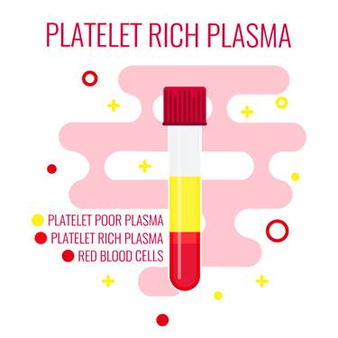
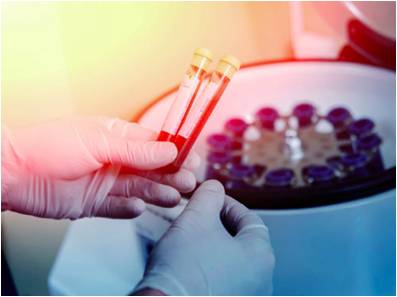
In platelet-rich plasma prolotherapy, the platelet-rich plasma is extracted from the patient under sterile conditions in clinical settings. This blood is then centrifuged and different components of the blood are separated into different layers. The platelet-rich plasma layer is separated and the rest of the material is discarded. The platelet-rich plasma is injected at the site of injury. When the platelet-rich plasma is injected at the site of injury, it releases alpha granules that cause the release of one' own growth factors. Growth factors are the proteins that initiate the tissue repair by cell growth and proliferation. Platelets cause the growth factors to stimulate the epithelial growth factors (EGF) which induce the cell migration and replication at the site of damage, causing the damaged tissues to heal quickly.
The platelet-rich plasma injection is followed by three stages of healing:
- Inflammation phase that lasts for 2-3 days. In this phase, growth factors are released.
- Proliferation phase that lasts for 2-4 weeks. It is vital for musculoskeletal regeneration.
- Remodeling which lasts over a year. In this phase, collagen is matured and strengthened.
The platelet-rich plasma Prolotherapy specialist makes sure that these three phases are completed to ensure the complete recovery from the injury.
Safety IssuesThe main safety concerns when using an invasive method are:
- Immunogenic reaction
- Disease transfer
- Hyperplasia
- Cancer
- Tumour growth
Platelet-rich plasma Prolotherapy uses the blood of the patient thus there are no chances of immunogenic reaction or blood-related disease transfer. No studies have documented that platelet-rich plasma Prolotherapy causes tumor growth or cancer.
The symptoms are temporarily worse after the injection. The possible side effects of platelet-rich plasma Prolotherapy are:
- Infection
- No relief of pain
- Neurovascular injury
- Scar tissue formation
- Loss of limb or death is rare, but possible

Recent evidence shows that PRP prolotherapy can be helpful in treating foot conditions like patellar tendinopathy. Prolotherapy was seen to improve ligament strength in a patient who was suffering from a painful foot and was unresponsive to NSAIDs.
10% of the patients with plantar vasculopathy are nonresponsive to conventional treatment options. In a detailed review of PRP prolotherapy effectiveness, the researchers found great improvement in the symptoms at the baseline and during the follow-up assessments. The success rate of Plantar vasculopathy was 66.6% in the patients after 6 months.
The effectiveness of PRP prolotherapy is evaluated by preclinical and human cell culture studies. The studies have shown increased tendon repair without scar formation after PRP injections. Another study showed a 60% improvement in the patients treated with platelet-rich plasma Prolotherapy for damaged tendons.
The effectiveness of the PRP is also evaluated for muscle damage repair. Although limited studies are available that show the effectiveness of this technique in muscle damage repair, experiments done in the lab on the human cells have shown the effectiveness of this technique for muscle repair and cell growth.
Surgery remains the last option when all the conventional treatment options fail, but it involves the risk of nerve injury and infection. PRP Prolotherapy has shown no side effects in more than 1,000 patients treated with PRP injections for various conditions.
PRP Prolotherapy is an effective treatment option. Several pieces of evidence are available, but they are limited. There is a need to conduct a full clinical trial to establish the effectiveness of PRP prolotherapy.

While PRP and stem cell treatments are enhancing the tissue repair and regeneration, conservative treatments can enhance healing, strengthen the muscles, and stabilize joint movements to maximize your recovery.
COLD LASER THERAPY TREATMENTS
- Accelerated tissue repair and cell growth
- Faster wound healing
- Reduced fibrous tissue formation
- Anti-inflammation
- Pain relief
- Increased blood flow
- Increased repair and regeneration
- Nerve function and repair
- Increased energy production- ATP
Photons of light from lasers penetrate into tissue and accelerate cellular growth and reproduction. Laser therapy increases the energy available to the cell so it can work faster, better, and quickly get rid of waste products. When cells of tendons, ligaments, and muscles are exposed to laser light they repair and heal faster.

Laser light increases collagen production by stimulating fibroblasts. Collagen is the building block of tissue repair and healing. Laser therapy increases fibroblast activity and therefore collagen production to speed healing.
Low-level laser therapy decreases scar tissue formation. Scar tissue can be a source of chronic pain and poor healing. By eliminating excessive scar tissue and encouraging proper collagen production, painful scars and chronic pain is reduced.
Laser therapy causes vasodilatation (increases the size of capillaries) which increases blood flow. The treatments also increases lymphatic drainage to decrease swelling or edema. Therefore, laser therapy reduces swelling caused by bruising or inflammation while speeding the recovery process.
Cold laser therapy decreases pain by blocking pain signals to the brain. Some nerve cells sense pain and send signals to the brain. Chronic pain can be caused by overly active pain nerves. Specific wavelengths help "shut off" the pain signals, thereby eliminating your pain.
Low-level lasers are excellent at decreasing inflammation, which also increases pain nerve activity. Cold laser therapy also increases endorphins and enkephalins, which block pain signals and decrease pain sensation. Overall laser therapy reduces painful nerve signals and reduces your perceived pain.

Blood carries nutrients and building blocks to the tissue, and carries waste products away. Increased blood flow to tissues increases and enhances cellular healing. Cold laser therapy increases the formation of capillaries in damaged tissue. Specific laser frequency also increases blood flow to the area treated to enhance injury repair.
Low-level lasers increases enzyme activity to improve metabolic activity that affects cell repair and regeneration. The enzymes are turned on "high" to speed the healing.
Nerves heal very slowly. Lasers speed up this process. Damage to nerves causes numbness, pain, muscle weakness, and altered sensations. Laser therapy treatments enhance nerve function, healing, and reduce pain.
ATP is like gasoline for cells, it is the energy source that cells operate. Injured cells often have low levels of ATP, which decreases their ability to heal and repair. By increasing ATP and "gasoline storage levels," cells have the ability to heal and repair.

Therapeutic treatments for addressing soft tissue injuries involve massage therapy, manual therapy, trigger point therapy, Graston Technique, or Active Release Technique. These treatments increase blood flow, decrease muscle spasms, enhance flexibility, speed healing, and promote proper tissue repair.
When these treatments are incorporated into a treatment plan, patients heal faster and are less likely to have long-term pain, soft tissue fibrosis, or scar tissue in the injured muscle. These soft tissue treatments are incorporated with therapeutic exercises and flexibility programs.
Many leg injuries are associated with radiating pain. The two legs function as a system for movement. Injuries in one area of the system are commonly associated with poor joint stabilization in the foot, knee, or hip. This leads to poor alignment and excessive forces being placed onto muscles and tendons. Knee injuries are common because of weakness and poor stabilization of the leg and hip muscles. The combination of muscle weakness, poor coordination, and altered gait mechanics produce excessive strain on the soft tissues.
The lower extremities work as a comprehensive unit performing many of the repetitive tasks at home, work, and recreational sports. Injuries to one area of the musculature often indicate that additional damage has been incurred by other muscles.

Many therapeutic exercises can help restore proper strength and endurance to the leg muscles. Isometric exercises are often the initial treatment exercises, followed by single plane rubber band exercises for hip, knee, and ankle; flexion, extension, adduction, abduction, circumduction, inversion, and eversion. Dynamic exercises involving stability foam, rubber discs, exercise balls, and BOSU balls can be performed on the floor. The more unstable of the surface the more effort and stabilization is required of all the lower extremity muscles.
Vibration plates enhance neuromuscular learning throughout the ankle, knee, foot, hip, and back muscles. Additional strength exercises can be found on the hip, knee, and foot strengthening pages. More information for injuries and treatments foot pain and exercises.
BIBLIOGRAPHY
Franceschi, F., Papalia, R., Franceschetti, E., Paciotti, M., Maffulli, N., & Denaro, V. (2014). Platelet-rich plasma injections for chronic plantar fasciopathy: a systematic review. Br Med Bull, 83-95.
Rabago, D., Slattengren, A., & Zgierska, A. (2010). Prolotherapy in Primary Care Practice. Prim Care, 65–80.