
Chiropractor Chandler AZ
PLATELET RICH PLASMA PROLOTHERAPY FOR CHONDROMALACIA PATELLA
Chondromalacia is a condition in which the articular cartilage softens and wears out. The patella is the kneecap which covers the knee joint. Chondromalacia patella is a condition in which the articular cartilage on the undersurface of the kneecap is softened and then deteriorated. This condition is also known as runner's knee. It is more common in young athletes, but elderly suffering from arthritis can also get affected by it. It is the most common cause of knee pain in the younger population.
CAUSES OF CHONDROMALACIA PATELLA:
The actual cause of Chondromalacia patella is unknown, but it is considered to be an injury caused by overuse of the knee. According to researchers, the following can cause Chondromalacia patella:
- Q-angle: The most important cause of Chondromalacia patella is abnormal Q-angle. Normal Q-angle for men is 14◦ and for women, it is 17◦. The lateral pull of the patella is increased if this angle is increased which then can lead to Chondromalacia patella.
- Muscle tightness: If the rectus femoris muscle is tightened, it affects the patellar movement when the knee is bent. Iliotibial band muscle tightness pulls the patella to the side of the knee when the knee is bent.
- Trauma: Knee instability caused by a previous trauma or misuse of the knee can lead to Chondromalacia patella. Repetitive trauma and inflammatory conditions like arthritis can increase the chances of Chondromalacia patella.
There are certain factors that increase the risk of Chondromalacia patella such as:
- Age: the young population is at higher risk of Chondromalacia patella. During this age, the bones and muscles are developing very fast which may cause short-term muscle imbalance.
- Gender: women have less muscle mass as compared to men and that is why they are more likely to suffer from Chondromalacia patella. Less muscle mass is the cause of abnormal knee positioning and more pressure on the patella.
- Flat feet: people with flat feet are at higher risk of Chondromalacia patella as the flat feet put more pressure on the knee joint.
- Previous injury: any previous knee injury such as dislocation can enhance the chances of Chondromalacia patella.
- High activity level: People who have high activity level or participate in activities that involve excessive running or jumping are at higher risk of Chondromalacia patella.
- Arthritis: Chondromalacia patella is sometimes a symptom of arthritis which prevents the patella from functioning properly.

There are 4 grades of Chondromalacia patella based on the severity of the disease. Grade 1 is an indicator of less severe Chondromalacia patella while grade 4 shows the highest severity level.
- Grade 1: indicates softening of the articular cartilage under the patella.
- Grade 2: indicates softening of articular cartilage with anomalous surface characteristics.
- Grade 3: indicates thinning of articular cartilage with tissue degradation.
- Grade 4: the most severe stage that indicates the bone exposure with a high level of articular cartilage degradation.
The symptoms of Chondromalacia patella are different from osteoarthritis. These symptoms include:
- Only one knee is affected
- Localized softening and degradation of articular cartilage in the knee
- Increase in pain after daily activities like running, jumping, climbing stairs
- Tenderness
- Decrease in quadriceps strength
- Cracking sensation in the knee
Diagnosis of Chondromalacia patella is often difficult because its actual cause is still unknown. There are different ways to diagnose this condition, however.
EXAMINATIONKnee examination is divided into 3 sections:
- Look: The knee joint looks normal in most of the cases but in some cases, there might be a minor effusion that can be an indication of Chondromalacia patella.
- Feel: Pain when the patella is rubbed again femur.
- Move: repetitive knee movements will be painful.
- X-ray: X-ray does not provide any information when Chondromalacia patella is at the early stages, but in advance stages, patellofemoral joint space tapers are visible on X-rays.
- Pinhole scintigraphy: It is a form of arthrography that is helpful in the diagnosis of Chondromalacia patella.
- MRI: It is an efficient way to diagnose Chondromalacia patella. It is non-invasive and easy to perform. The benefit of this test is that sensitivity and specificity of the diagnosis can be increased by increasing the strength of the magnets and mounting a specialized coil.
The goal of treatment is to decrease the pressure on the patella and knee. Rest, stabilizing the knee, and icing are the first line of treatment. Cartilage can repair itself in most Chondromalacia patella cases.
MEDICATIONIf the first line of treatment fails, the doctor will prescribe anti-inflammatory medicines such as Ibuprofen to reduce the inflammation and pain in the affected knee. If inflammation and pain persist, then the doctor will suggest the following treatment options:
PHYSICAL THERAPYThe main focus of physical therapy is to strengthen quadriceps, hamstrings, adductors and abductors to improve muscle strength and balance. It will avoid knee imbalance. Light exercises that do not involve weights, such as swimming, are also recommended.
SURGERYIf the physical therapy fails to relieve the pain and inflammation, surgery is the only option left. Arthroscopic surgery is performed to examine the knee joint and determine if there is a knee alignment problem. Another common procedure is the lateral release which involves cutting some of the ligaments to release tension.
For more information about Chondromalacia Patella, please visit here
ALTERNATIVE TREATMENT OPTION
Chondromalacia patella is characterized by cartilage damage. The current treatment options do not repair the cartilage, but rather they try to relieve the pain by reducing inflammation and giving the cartilage a chance to repair itself. Any treatment option that can accelerate and assist in the process of self-repair will increase the repair and decrease the recovery time. Platelet-rich plasma (PRP) prolotherapy is a new procedure that works by using the repair machinery of the body. PRP prolotherapy is a part of a new branch of medicine known as Regenerative Medicine. It uses the body's healing ability to repair the damage and relieve the pain and inflammation. The platelet-rich plasma Prolotherapy works on a very simple principle: "when the platelet concentration is increased in a certain area of the body, it accelerates the healing process." Platelets contain many chemicals known as:
- Glycogen
- Lysosomes
- Alpha granules
- Beta granules
Alpha granules contain growth factors and they are the main focus of platelet-rich plasma therapy. There are three stages of healing after platelet-rich plasma Prolotherapy injection and different types of growth factors are involved in driving the different stages:
- Inflammation phase: It lasts for 2-3 days.
- Proliferation phase: It lasts for 2-4 weeks.
- Remodeling phase: It lasts over a year. In this phase, collagen is matured and strengthened and injury is healed.
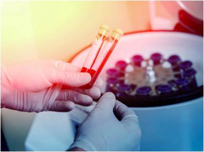
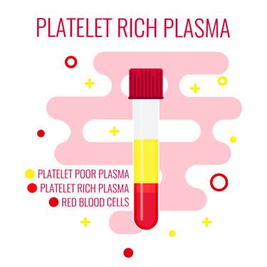
20-30 mL of blood is used to make platelet-rich plasma. The blood is spun for 15 minutes at 3,200 rpm in a centrifuge machine. This step separates platelet-rich plasma from platelet-poor plasma. 3 mL of platelet-rich plasma is obtained from 20 mL of blood. The platelet-rich plasma is neutralized by adding half mL of sodium bicarbonate that neutralizes its pH. The injection is administered with the help of dynamic musculoskeletal ultrasound with a transducer of 6–13 Hz to maximize the location accuracy for the injection. The injection is administered directly at the site of injury.
As platelet-rich plasma Prolotherapy technique uses the patient's own blood, the chances of immunogenic reaction or transfer of blood borne diseases are completely eliminated.
The growth factors attach to the cell surface and not the nucleus of the cell, thus the chances of tumor growth are also eliminated. As the platelet-rich plasma Prolotherapy is an injection-based procedure, the risks involved in are:
- Anaesthesia allergy
- Infection
- Neural trauma
- Organ trauma
- Needle breakage
Some data is available that shows the effectiveness of PRP prolotherapy in treating Chondromalacia patella. A research was done on 117 patients in 2007-2008 that had Chondromalacia patella. The results of the research showed that PRP prolotherapy had improved knee functionality, range of motion, and decreased pain. PRP injection also improved Chondromalacia patella symptoms in all patients. 92% of patients showed improvement in pain and the range of motion and stiffness. 96% of patients in this study said that prolotherapy has improved their life.
PRP Prolotherapy repairs the structure of the patella by activating and accelerating the body's healing machinery. Thus it is thought to protect the joint along with providing pain relief.
The data available on prolotherapy shows that PRP prolotherapy is safe and has fewer to no side effects.
In the U.S, the cost of RPR treatment varies. In Florida, a single PRP injection for knee pain costs $800. In New York, cost varies from $1000-$2000 per injection.
PRP prolotherapy for Chondromalacia patella is a very promising option that can help in pain relief and increase the functionality of the knee. It can be considered a first-line conservative therapy for Chondromalacia patella but a large, multicentre randomized trial study is needed to further assess the efficacy of PRP treatment for patients with Chondromalacia patella.
CONSERVATIVE TREATMENTS TO COMBINE WITH PRPWhile PRP and stem cell treatments are enhancing the tissue repair and regeneration, conservative treatments can enhance healing, strengthen the muscles, and stabilize joint movements to maximize your recovery.
COLD LASER THERAPY TREATMENTS
- Accelerated tissue repair and cell growth
- Faster wound healing
- Reduced fibrous tissue formation
- Anti-inflammation
- Pain relief
- Increased blood flow
- Increased repair and regeneration
- Nerve function and repair
- Increased energy production- ATP
Photons of light from lasers penetrate into tissue and accelerate cellular growth and reproduction. Laser therapy increases the energy available to the cell so it can work faster, better, and quickly get rid of waste products. When cells of tendons, ligaments, and muscles are exposed to laser light they repair and heal faster.

Laser light increases collagen production by stimulating fibroblasts. Collagen is the building block of tissue repair and healing. Laser therapy increases fibroblast activity and therefore collagen production to speed healing.
Low-level laser therapy decreases scar tissue formation. Scar tissue can be a source of chronic pain and poor healing. By eliminating excessive scar tissue and encouraging proper collagen production, painful scars and chronic pain is reduced.
Laser therapy causes vasodilatation (increases the size of capillaries) which increases blood flow. The treatments also increases lymphatic drainage to decrease swelling or edema. Therefore, laser therapy reduces swelling caused by bruising or inflammation while speeding the recovery process.
Cold laser therapy decreases pain by blocking pain signals to the brain. Some nerve cells sense pain and send signals to the brain. Chronic pain can be caused by overly active pain nerves. Specific wavelengths help "shut off" the pain signals, thereby eliminating your pain.
Low-level lasers are excellent at decreasing inflammation, which also increases pain nerve activity. Cold laser therapy also increases endorphins and enkephalins, which block pain signals and decrease pain sensation. Overall laser therapy reduces painful nerve signals and reduces your perceived pain.
Blood carries nutrients and building blocks to the tissue, and carries waste products away. Increased blood flow to tissues increases and enhances cellular healing. Cold laser therapy increases the formation of capillaries in damaged tissue. Specific laser frequency also increases blood flow to the area treated to enhance injury repair.
Low-level lasers increases enzyme activity to improve metabolic activity that affects cell repair and regeneration. The enzymes are turned on "high" to speed the healing.
Nerves heal very slowly. Lasers speed up this process. Damage to nerves causes numbness, pain, muscle weakness, and altered sensations. Laser therapy treatments enhance nerve function, healing, and reduce pain.
ATP is like gasoline for cells, it is the energy source that cells operate. Injured cells often have low levels of ATP, which decreases their ability to heal and repair. By increasing ATP and "gasoline storage levels," cells have the ability to heal and repair.
Therapeutic treatments for addressing soft tissue injuries involve massage therapy, manual therapy, trigger point therapy, Graston Technique, or Active Release Technique. These treatments increase blood flow, decrease muscle spasms, enhance flexibility, speed healing, and promote proper tissue repair.
When these treatments are incorporated into a treatment plan, patients heal faster and are less likely to have long-term pain, soft tissue fibrosis, or scar tissue in the injured muscle. These soft tissue treatments are incorporated with therapeutic exercises and flexibility programs.
Many leg injuries are associated with radiating pain. The two legs function as a system for movement. Injuries in one area of the system are commonly associated with poor joint stabilization in the foot, knee, or hip. This leads to poor alignment and excessive forces being placed onto muscles and tendons. Knee injuries are common because of weakness and poor stabilization of the leg and hip muscles. The combination of muscle weakness, poor coordination, and altered gait mechanics produce excessive strain on the soft tissues.
The lower extremities work as a comprehensive unit performing many of the repetitive tasks at home, work, and recreational sports. Injuries to one area of the musculature often indicate that additional damage has been incurred by other muscles.

Many therapeutic exercises can help restore proper strength and endurance to the leg muscles. Isometric exercises are often the initial treatment exercises, followed by single plane rubber band exercises for hip, knee, and ankle; flexion, extension, adduction, abduction, circumduction, inversion, and eversion. Dynamic exercises involving stability foam, rubber discs, exercise balls, and BOSU balls can be performed on the floor. The more unstable of the surface the more effort and stabilization is required of all the lower extremity muscles.
Vibration plates enhance neuromuscular learning throughout the ankle, knee, foot, hip, and back muscles. Additional strength exercises can be found on the hip, knee, and foot strengthening pages. More information for injuries and treatments foot pain and exercises.
BIBLIOGRAPHY:
Hauser, R. A., & Sprague, I. S. (2014). Outcomes of Prolotherapy in Chondromalacia Patella Patients: Improvements in Pain Level and Function. Clin Med Insights Arthritis Musculoskelet Disord, 13–20.