
Chiropractor Chandler AZ
Upper Crossed Syndrome: Diagnosis and Treatment
Upper Crossed Syndrome is a term first used by the Czech physician, Vladimir Janda, to describe a group of muscle imbalances often seen in patients with rounded shoulders and forward neck flexion. Because so much of today’s working population uses computers, and because many of these individuals are sedentary for most if not all of their waking hours, this syndrome and its complications ranging from upper back pain to cervicogenic headaches are becoming more and more common Moore, 2004). In addition, as children and adolescents spend much of their time on mobile phones and tablets, they are developing some of these symptoms earlier in life than their parents.
Janda used the term, “upper crossed,” because the weakened and shortened muscles involved in this syndrome cross in the mid-back. In addition to upper back, head and neck pain, upper crossed syndrome can lead to shoulder injuries such as impingement, degeneration of the supraspinatus and other rotator cuff tendons, and lower back pain, since many of these individuals develop a compensatory posterior pelvic tilt.
Although studies of upper crossed syndrome have utilized X-ray imaging and EMG studies to confirm observations, for the purpose of treatment, your assessment can be much simpler. Beginning with standing posture, looking for weak deep neck flexors, overactivated sternocleidomastoid muscles, overactivated upper trapezius, scalenes and levator scapulae, weakened and elongated middle and lower trapezius, tight pectoral and weakened serratus anterior muscles. Put simply, the muscles at the base of the back of the neck are shortened and overactive, overactivated SCM muscles are compensating for weakened deep neck flexors, and both chest muscles and the anterior shoulder capsule are tight and internally rotated (Arshadi et al., 2019).
If you ask the patient to abduct his/her arms, you will likely see that the scapulae are protracted and possibly winged (due to weak serratus anterior and lower trapezius muscles). Because many persons with upper crossed syndrome are sedentary, other important back muscles such as the rhomboids and lateralis are weak as well. As a result, these large muscles fail to assist with postural stabilization.

Chiropractic Adjustment and Corrective Exercise
Studies on both older working and college-age adults have shown chiropractic adjustment and corrective exercise to be efficacious in resolving this constellation of symptoms (Arshadi et al., 2019). In fact, a case study on a 56-year-old patient with severe cervicogenic headaches confirmed that chiropractic adjustment, trigger point myofascial massage and corrective exercise was more effective both short and long-term in resolving symptoms than pharmacotherapy (Moore, 2004).
In our clinic, we utilize a combination of conservative treatment modalities including chiropractic adjustment, shockwave, cold laser, interferential electrotherapy, ice, stretching and corrective exercise to treat the various symptoms of upper crossed syndrome. Your starting point will depend on the patient’s age and history of physical activity (or lack thereof), as well as other medical conditions that might be contributing factors. At a point in time when a high percentage of the US population is being diagnosed with diabetes and many individuals are also cancer survivors, it is important to include a balance assessment as part of any diagnostic workup. Peripheral neuropathy, history of osteoarthritis and joint replacement surgeries can all contribute to the development of upper crossed syndrome.

Since the top priority is decreasing the patient’s pain level, begin with modalities directed towards that end: chiropractic adjustment to realign the cervical spine, myofascial release or shockwave to reduce tightness in the scalenes, levator scapulae and upper trapezius muscles, cold laser, interferential, ice and stretching for analgesia.
Get the patient started on a home therapy regimen of icing and stretching from the onset. This will help to control the discomfort and hasten the recuperation process. A simple “doorway stretch” (see photo) will begin to release tightness in the pectoral muscles and anterior shoulder capsules. Other shoulder stretches illustrated in this article require no special equipment, and will help to maintain flexibility gains made in your clinic. In addition, McKenzie exercises that enhance flexibility and reduce pain are easy to perform at home (McKenzie, 2011).
In order to improve postural stability, you will need to teach the patient what proper posture feels like. Begin by photographic the patient from the side, so he/she can see the extent of forward neck flexion and inwardly rotated shoulders. In some patients, you may also observe hyper-kyphosis. Chin tuck exercises are one way to reduce overactivated SCMs and reactivate the deep neck flexor muscles.
We also teach our patients to “pack their scapulae.” Begin by placing your hands on the patient’s shoulder blades, to give them a tactile reference. Keeping your hands on the shoulder blades, have the person focus on lowering his/her shoulders and pushing the shoulder blades together until the medial edges of the scapulae are parallel and a couple of inches apart. Those individuals with severe winging and/or protraction may need to practice in order to move their scapulae into the proper position. A spouse or friend can assist with the tactile reference, so the patient can practice this at home.
Where you begin with strengthening exercises depends on the patient’s symptoms. If the individual has low back pain, we recommend beginning with core exercises to activate the glutes and abdominal muscles. If the pain is localized to the upper back and neck, with or without headaches, begin with the scapular stabilizers.
Core Strengthening
The term, “core,” is poorly defined and can mean different things depending upon the context. Here we are referring to the three sets of gluteal muscles, abdominal muscles (including obliques) and spinal extensors. Begin with a basic bridging exercise with the patient supine on a treatment table. The progression begins with feet and knees shoulder width apart, then feet and knees together and finally single leg bridging. Once the patient is strong enough to perform unipedal bridges he/she can progress to bridging on an exercise ball. Bridging works all three sets of glutes, so it is a good way to build pelvic stability for walking as well as creating a stable base for the middle back muscles.
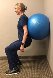
Leg circles are an alternate exercise for gluteus medius and minimus muscles. Rather than have patients perform classic standing squats, we recommend ball wall squats as a safer alternative, particularly for older adults.

Planks (prone and lateral) build strength through both the abdomen and gluteal muscles. For patients who are less fit, begin with either a wall plank (easiest) or by having the individual rest his/her arms on an elevated bench or table. Patients with weak core muscles will tend to ‘cheat’ by activating the same tight shoulder muscles you are trying to lengthen, so watch for proper form (no hunched shoulders).

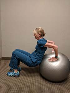
Tricep dips off a bench or for more advanced patients, utilizing an exercise ball are an alternative for activating all of the core muscles in a dynamic manner.

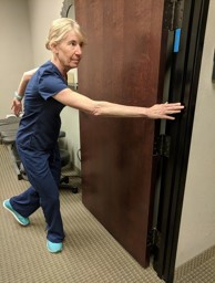
The door frame touch exercise (see photo) activates the glutes and helps patients build balance during transverse movements.
Middle Back Strengthening
Exercises for the middle back include front and side lat raises (light weight with high reps), upright rows, tricep kickbacks, forward and reverse rows. Patients can do these at home by either purchasing some inexpensive light dumbbells (2-5 lbs), or using small water bottles. Therabands can be tied to a railing or door knob to perform rows. As an alternative, inexpensive cables are typically available at athletic supply stores.
For the arms, have your patient perform sets of external rotation using a Theraband loop (see photo).
Warm-up and Cool-down
All exercise sessions should begin with a five minute warm-up and conclude with five minutes of cooling down.
Since static stretching temporarily decreases muscle strength, the warm-up should consist of dynamic stretches such as woodchoppers, dynamic balance exercises and walking lunges.
Save static stretches for the cool down, asking the patient to perform each stretch twice and hold the position for 30 seconds.
References
Arshadi, R., Ghasemi, G. & Samadi, H. (2019). Effects of an 8-week selective corrective exercises program on electromyography activity of scapular and neck muscles in persons with upper crossed syndrome: Randomized controlled trial. Physical Therapy in Sport, 37, 113-119. https://doi.org.10.1016/j.ptsp.2019.03.008
McKenzie, R. (2011). Treat Your Own Neck (5th Ed.). Orthopedic Physical Therapy Products.
Moore, M. (2004). Upper Crossed Syndrome and its Relationship to Cervicogenic Headache. Journal of Manipulative and Physiological Therapeutics, 27, 414-420. https://doi.org.10.1016.jmpt.2004.05.007